
In modern digital dentistry, intraoral dental X-ray sensors have become indispensable for diagnosis, treatment planning, and patient communication. Whether detecting early caries, evaluating root canal treatments, or assessing bone conditions, image quality directly affects clinical accuracy.
Many dental professionals ask the same questions:
- How does a dental sensor work?
- What is the difference between CCD and CMOS sensors?
- How long should a dental sensor last?
- When is it time to replace an aging sensor?
This article explains the working principle of dental sensors, compares CCD and CMOS technologies, and provides practical guidance on sensor lifespan and replacement.
How Does a Dental X-ray Sensor Work?
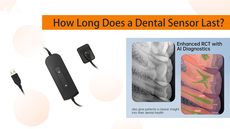
Although different sensor technologies exist, almost all modern dental intraoral sensors follow the same imaging process.
Step 1. X-rays Pass Through the Tooth
During an examination, the X-ray beam penetrates the patient’s teeth and surrounding bone. Different tissues absorb different amounts of radiation, creating varying X-ray intensities.
Step 2. The Scintillator Converts X-rays into Visible Light
The first layer inside the sensor is the scintillator (fluorescent layer).
Its job is to convert invisible X-rays into visible light that the imaging chip can detect.
This layer is one of the most critical components of the entire sensor because it largely determines image brightness, sharpness, and contrast.
Step 3. The CMOS Imaging Chip Captures the Light
The visible light reaches millions of microscopic pixels on the CMOS imaging chip.
Each pixel converts light into an electrical charge.
Step 4. Digital Processing Creates the Image
The electrical signals are processed electronically and transferred to the imaging software.
Within seconds, the clinician receives a grayscale dental image that can reveal:
- Dental caries
- Periapical lesions
- Root canal anatomy
- Periodontal bone loss
- Trabecular bone structure
- Impacted teeth
CCD vs CMOS: What’s the Difference?
Years ago, dental sensors mainly used either CCD (Charge-Coupled Device) or CMOS (Complementary Metal-Oxide Semiconductor) technology.
Interestingly, both technologies produce images using the exact same X-ray conversion process. The only major difference is how the electrical charges are read from the sensor.
| Feature | CCD | CMOS |
|---|---|---|
| Manufacturing Cost | Higher | Lower |
| Production Process | Complex | Standard semiconductor process |
| Power Consumption | Higher | Lower |
| Image Speed | Slower | Faster |
| Dynamic Range | Traditionally excellent | Modern back-illuminated CMOS matches or exceeds CCD |
| Typical Applications | Scientific imaging, broadcasting, astronomy | Smartphones, digital cameras, medical imaging, automotive cameras, dental sensors |
Why Has CMOS Become the Industry Standard?
Twenty years ago, CCD offered superior image quality.
However, advances in backside-illuminated (BSI) CMOS technology have dramatically improved performance.
Today’s CMOS sensors provide:
- Higher imaging speed
- Lower power consumption
- Better manufacturing consistency
- Reduced production costs
- Excellent image quality
- Greater durability
As a result, nearly all modern dental intraoral sensors now use CMOS technology.
CCD technology remains primarily in specialized scientific research and certain high-end industrial imaging systems.
What Determines the Lifespan of a Dental Sensor?
Many people assume that the CMOS chip eventually wears out.
In reality, the imaging chip is rarely the first component to fail.
The Scintillator Determines Sensor Life
The scintillator layer is responsible for approximately 80% of long-term image quality degradation.
Over years of daily use, several factors gradually reduce its performance.
1. Continuous X-ray Exposure
Repeated X-ray exposure slowly ages the phosphor material.
As light conversion efficiency decreases, images become:
- Less bright
- Lower in contrast
- More difficult to interpret
2. Chemical Disinfection
Routine cleaning and disinfection can gradually affect the sensor’s protective surface.
Over time, disinfectants may contribute to:
- Localized cloudiness
- Image haze
- Permanent dark spots
Proper barrier protection and manufacturer-recommended cleaning procedures can help minimize this risk.
3. Mechanical Stress
Dental sensors experience significant physical stress during daily use.
Repeated biting pressure, accidental drops, or impacts may permanently damage the scintillator layer, leading to:
- Fixed image artifacts
- Dead areas
- Permanent dark marks
Does the CMOS Chip Wear Out?
Under normal clinical conditions, CMOS chips are extremely durable.
The electronic components are typically capable of functioning for 8–10 years or longer before experiencing noticeable electrical degradation.
In most cases, image quality declines well before the chip itself reaches the end of its life, primarily because the scintillator layer deteriorates first.
Expected Lifespan in a Standard Dental Practice
For a general dental clinic taking approximately 10–20 radiographs per day, image quality usually follows this pattern:
Years 1–5
- Stable image quality
- Excellent contrast
- No noticeable increase in radiation exposure
- Consistent diagnostic performance
Years 5–7
Gradual image degradation begins.
Clinicians may observe:
- Slight image haze
- Increased image noise
- Reduced sharpness
- Minor increases in exposure settings to maintain image quality
After 7 Years
Image deterioration becomes clinically significant.
Common problems include:
- Fixed dark spots
- Vertical or horizontal lines
- Reduced local sensitivity
- Poor visualization of fine root canal anatomy
- Difficulty detecting small carious lesions
For most general practices, replacement around seven years is considered appropriate.
Lifespan in High-Volume Clinics
High-volume practices such as:
- Orthodontic centers
- Multi-chair clinics
- Large group practices
often capture 30–60 radiographs per day.
Because the scintillator receives much heavier cumulative radiation exposure, image degradation occurs sooner.
Typical expectations are:
- Noticeable quality decline after approximately 3.5–5 years
- Replacement recommended within 5–6 years
Theoretical Image Count vs Practical Service Life
Manufacturers often estimate dental sensors for approximately 100,000 exposures under ideal operating conditions.
Based on daily usage:
| Daily Images | Estimated Exposure Life |
|---|---|
| 30 images/day | Approximately 9 years |
| 60 images/day | Approximately 4.5 years |
However, medical device manufacturers generally specify a design service life of about 7 years, with a practical replacement window between 5 and 8 years, depending on workload and maintenance.
Clinical image quality—not exposure count alone—should guide replacement decisions.
Signs That Your Dental Sensor Should Be Replaced
Aging sensors often display recognizable symptoms.
Consider replacing the sensor if you notice:
- Images becoming increasingly hazy under the same exposure settings
- Poor contrast and blurred trabecular bone structure
- Permanent dark spots or cloudy areas
- Vertical or horizontal lines that calibration cannot remove
- Increasing radiation exposure needed to achieve acceptable images
- Excessive noise in low-dose imaging modes
- Difficulty visualizing fine root canal details or early caries
When these issues persist, continued use may compromise diagnostic confidence.
Best Practices to Extend Sensor Life
Although no sensor lasts forever, proper care can significantly extend its service life.
Recommended practices include:
- Use disposable protective barriers for every patient.
- Follow the manufacturer’s approved disinfection procedures.
- Avoid excessive bending or twisting of the sensor cable.
- Prevent patients from biting directly on sensor edges whenever possible.
- Store the sensor in a protective holder when not in use.
- Handle the sensor carefully to minimize accidental drops or impacts.
Recommended Replacement Timeline
The following schedule provides a practical reference for most clinics:
| Clinic Type | Daily Radiographs | Recommended Replacement |
|---|---|---|
| General Dental Practice | 10–20 | Around 7 years |
| High-Volume Clinic | 30–60 | Around 5–6 years |
Routine evaluation of image quality should always accompany these general guidelines.
Final Thoughts
Modern CMOS dental sensors offer outstanding image quality, reliability, and efficiency, making them the standard choice for today’s dental practices. While the CMOS chip itself is designed for long-term performance, the scintillator layer is typically the first component to age, gradually reducing image clarity after years of clinical use.
For most dental clinics, replacing an intraoral sensor after about seven years helps maintain diagnostic accuracy. High-volume practices may benefit from replacement after five to six years, depending on daily workload and image quality.
By understanding how dental sensors work and monitoring signs of image degradation, clinics can ensure consistent diagnostic performance while optimizing both patient safety and clinical efficiency.
Post time: Jul-25-2026