
Digital dentistry is changing how dental professionals diagnose oral conditions, plan treatments, and manage complex procedures. Among the most important technologies driving this change is Dental Cone Beam Computed Tomography (CBCT).
Unlike conventional 2D dental X-rays, a dental CBCT scanner captures three-dimensional images of teeth, jawbones, and surrounding anatomical structures. This allows dentists to examine areas from different angles and obtain more detailed information before treatment.
With the development of artificial intelligence (AI), modern dental CBCT systems are moving beyond image acquisition. AI-assisted image analysis can help identify anatomical structures, perform measurements, and organize diagnostic information, supporting more efficient clinical workflows.
For dental clinics, implant centers, and hospitals, choosing an AI-powered dental CBCT scanner is becoming an important investment in digital imaging capabilities.
1. Why Is Dental CBCT Important for Modern Implant Planning?
Dental implant treatment requires careful evaluation of bone structure and surrounding anatomical areas.
Traditional panoramic X-rays provide a two-dimensional overview, but they cannot fully demonstrate the buccolingual dimensions of the jawbone or the three-dimensional relationship between an implant site and nearby anatomical structures.
CBCT imaging provides additional information for:
- Bone height and width evaluation.
- Bone volume assessment.
- Inferior alveolar nerve canal visualization.
- Maxillary sinus evaluation.
- Implant position and angulation planning.
- Assessment of anatomical limitations.
These details help clinicians evaluate implant sites and plan appropriate treatment approaches.
CBCT is particularly valuable when three-dimensional information is expected to influence clinical decisions. The examination should always be justified according to the patient’s individual clinical needs.
2. How Does AI Improve Dental CBCT Image Analysis?

Artificial intelligence is becoming an important development in dental imaging.
Traditional image analysis often requires dentists to manually identify anatomical landmarks, examine structures, and perform measurements. AI-assisted software can automate or support some of these processes.
Depending on the software functions available, AI-assisted CBCT analysis can support several tasks.
AI Nerve Canal Identification
Identifying the inferior alveolar nerve canal is an important part of mandibular implant planning.
AI-assisted nerve canal mapping can help dentists visualize the canal pathway and understand its relationship with the proposed implant site.
AI Tooth Segmentation
AI tooth segmentation helps separate individual teeth from surrounding anatomical structures in 3D images.
This can support dental analysis, treatment planning, and digital visualization.
AI Airway Analysis
Three-dimensional airway analysis is useful in selected orthodontic and maxillofacial assessment workflows.
AI-assisted segmentation can help visualize airway structures and provide additional information for professional evaluation.
AI Cephalometric Measurement
For orthodontic diagnosis, cephalometric analysis involves identifying anatomical landmarks and measuring craniofacial relationships.
AI-assisted landmark detection and measurement reports can help organize this process and reduce repetitive manual work.
Important: AI should be considered a clinical assistance tool, not a replacement for professional diagnosis. Dentists must review AI-generated results and make treatment decisions based on their clinical judgment.
3. 3-in-1 Dental Imaging: CBCT, PANO and CEPH in One System
Another important trend in digital dental imaging is multifunctional equipment integration.
Instead of purchasing separate imaging systems for different examinations, dental clinics can consider an integrated 3-in-1 dental imaging solution.
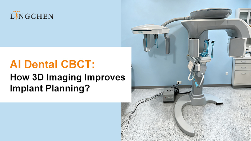
A modern CBCT system integrating CT, PANO, and CEPH provides three imaging modes.
CBCT / 3D CT Imaging
Provides three-dimensional information about teeth, jawbones, and anatomical structures.
Main applications include:
- Dental implant planning.
- Oral surgery evaluation.
- Jawbone assessment.
- Complex anatomical examination.
Panoramic Imaging (PANO)
Provides a two-dimensional overview of the upper and lower jaws.
Common applications include:
- General dental examinations.
- Tooth development assessment.
- Preliminary diagnosis.
- Overall dental structure evaluation.
Cephalometric Imaging (CEPH)
Supports orthodontic diagnosis through lateral cephalometric imaging and craniofacial measurements.
Common applications include:
- Orthodontic treatment planning.
- Facial skeletal assessment.
- Cephalometric measurements.
- Growth and development evaluation.
For dental clinics, hospitals, and universities, combining these imaging functions in one system can simplify equipment management, optimize room utilization, and support a wider range of diagnostic services.
4. What Should You Consider When Choosing a Dental CBCT Scanner?
Choosing a dental CBCT scanner involves more than comparing prices. Imaging specifications, clinical applications, software functions, and long-term operating requirements should all be evaluated.
Here are five important factors.
1. Field of View (FOV)
The field of view determines the anatomical area captured during scanning.
A larger FOV can support broader anatomical examinations, while a smaller FOV may be sufficient for localized clinical tasks.
For general dental practices and implant centers, a 16×9 cm FOV provides a useful range of imaging applications.
However, the appropriate FOV should always be selected according to the diagnostic requirement. The smallest FOV that adequately answers the clinical question should be preferred.
2. Voxel Size and Image Resolution
Voxel size is an important specification when comparing CBCT systems.
A smaller voxel size can provide finer spatial sampling, which may help visualize detailed anatomical structures.
For example, a CBCT scanner with a minimum voxel size of 0.05 mm (50 μm) is designed to capture detailed 3D dental images.
However, voxel size alone does not determine diagnostic image quality. Detector performance, exposure settings, patient movement, reconstruction algorithms, and imaging conditions also matter.
3. Detector Technology
The detector directly affects image acquisition and image quality.
CMOS detector technology is widely used in modern digital dental imaging systems.
When evaluating a CBCT scanner, buyers should consider detector performance, image consistency, noise characteristics, and clinical suitability.
4. AI Software and Image Analysis
AI-assisted functions can make image analysis more efficient.
Consider whether the system supports:
- AI nerve canal mapping.
- AI tooth segmentation.
- AI airway analysis.
- AI panoramic analysis.
- AI cephalometric landmark detection.
- Automatic measurement and report generation.
It is also important to check which functions are included, which require additional software, and whether the software is compatible with your existing digital workflow.
5. Scanning Speed and Clinical Workflow
Scanning efficiency is another important consideration for busy dental clinics.
A faster scanning process may help reduce examination time and patient movement.
However, scanning speed should not be evaluated separately from image quality, radiation exposure, and clinical diagnostic requirements.
Look for a system that balances acquisition efficiency with appropriate imaging protocols.
5. CBCT and Intraoral Scanners: Building a Digital Implant Workflow
One of the most important developments in digital dentistry is the integration of different imaging technologies.
CBCT and intraoral scanners provide different types of clinical information.
- CBCT provides 3D information about teeth, jawbones, and anatomical structures.
- Intraoral scanners capture detailed digital impressions of teeth and soft-tissue surfaces.
When these datasets are accurately registered using compatible planning software, clinicians can combine anatomical information with digital dental models.
A typical digital implant planning workflow includes:
- Acquire CBCT images of the patient’s oral and maxillofacial structures.
- Capture digital impressions using an intraoral scanner.
- Import both datasets into compatible planning software.
- Register the CBCT and intraoral scan data.
- Evaluate bone dimensions and anatomical structures.
- Plan implant position and angulation.
- Design a surgical guide when clinically indicated.
This workflow supports more comprehensive treatment planning and communication between clinicians, laboratories, and other digital dentistry professionals.
The accuracy of image registration, software compatibility, and professional verification remain essential throughout the process.
6. Radiation Safety: An Important Factor in Dental CBCT Selection
As dental CBCT becomes more widely used, radiation optimization remains an important consideration.
CBCT should not replace conventional dental radiography for every examination. Its use should be based on individual clinical indications and the additional diagnostic information required.
Dental professionals should consider:
- Selecting an appropriate field of view.
- Using exposure parameters suitable for the clinical task.
- Applying optimized imaging protocols.
- Avoiding unnecessary repeat examinations.
- Considering patient age and individual clinical requirements.
The objective is to obtain sufficient diagnostic information while keeping radiation exposure as low as reasonably achievable.
A high-quality dental CBCT solution should support appropriate clinical imaging protocols rather than simply prioritizing maximum image resolution.
7. Lingchen AI-Powered Dental CBCT Scanner: An Integrated 3-in-1 Imaging Solution

For dental clinics, implant centers, hospitals, and dental universities looking to develop their digital imaging capabilities, Lingchen offers an AI-powered dental CBCT scanner integrating three imaging modes in one system.
Its main specifications include:
| Feature | Specification |
|---|---|
| Imaging Technology | Cone Beam Computed Tomography |
| Detector | High-performance CMOS |
| Maximum FOV | 16×9 cm |
| Minimum Voxel Size | 0.05 mm |
| Imaging Modes | CT / PANO / CEPH |
| Fast Scan Technology | 3S UF Scanning |
| AI Functions | Intelligent AI-assisted analysis |
Key Features
AI-Assisted Image Analysis
Supports AI nerve canal mapping, tooth segmentation, jawbone segmentation, airway analysis, TMJ segmentation, panoramic analysis, and cephalometric measurements.
Three Imaging Modes in One System
CT, panoramic, and cephalometric imaging support different dental diagnostic and treatment planning requirements.
High-Resolution 3D Imaging
The CMOS detector, 16×9 cm maximum FOV, and 0.05 mm minimum voxel size provide detailed 3D imaging capabilities.
Applications Across Multiple Dental Specialties
Suitable for implant dentistry, orthodontics, oral surgery, and other dental imaging applications.
For clinics and institutions planning to upgrade their imaging facilities, an integrated system can provide a practical foundation for a more comprehensive digital dentistry workflow.
Learn more about the Lingchen AI-Powered Dental CBCT Scanner.
8. Frequently Asked Questions About Dental CBCT Scanners
What is the difference between dental CBCT and a panoramic X-ray?
A panoramic X-ray provides a two-dimensional image of the upper and lower jaws, while CBCT captures three-dimensional anatomical information.
CBCT is particularly useful when clinicians need to evaluate bone dimensions, nerve canal locations, and complex anatomical structures.
Is AI important in a dental CBCT scanner?
AI-assisted analysis can help identify anatomical structures, perform measurements, and organize imaging information. It can support clinical efficiency, but professional review and diagnosis remain essential.
What FOV is suitable for a dental CBCT scanner?
The appropriate FOV depends on the examination. A 16×9 cm FOV can support a range of implant, orthodontic, and oral diagnostic applications. Smaller FOV settings should be used when sufficient for the clinical question.
What is the difference between CBCT and CT?
Dental CBCT uses a cone-shaped X-ray beam and is specifically designed for dentomaxillofacial imaging. Conventional medical CT uses different acquisition technology and is designed for broader medical imaging applications.
The appropriate system depends on the clinical indication.
Can one CBCT scanner support implant and orthodontic applications?
Yes. A multifunctional system with CT, PANO, and CEPH imaging can support both implant planning and orthodontic diagnosis, subject to its imaging capabilities and clinical protocols.
Is a dental CBCT scanner suitable for universities and teaching hospitals?
Yes. CBCT systems can support dental education, clinical training, research, and diagnostic services. Universities should also consider installation requirements, radiation protection, room planning, software training, and after-sales support.
Conclusion
AI-assisted dental CBCT, 3D imaging, and integrated digital workflows are becoming important parts of modern dentistry.
When selecting a dental CBCT scanner, dental professionals should evaluate image quality, FOV, voxel size, detector technology, AI software, clinical applications, and radiation optimization together.
For dental clinics, implant centers, universities, and equipment distributors, an integrated CT, PANO, and CEPH imaging solution can provide a flexible foundation for expanding digital dental services.
Looking for an AI-powered dental CBCT scanner for your clinic, hospital, or distribution business?
Contact Lingchen Dental to learn more about our 3-in-1 dental CBCT imaging solution, technical specifications, and equipment supply options.
Post time: Nov-14-2026